Notes: Vision (Part 1)
(Part II of vision will come
from Brad's lecture)
10/26/12 - formatting change (no content change)
10/17/12 - Minor word editing
10/12/12 - Original posted version
Where we are going:
How is a distorted and upside-down 2-D retinal image transformed into the 3-D world we perceive?
Light
No species can see in the dark, but some are capable of seeing when there is little lightLight can be thought of as
- Particles of energy (photons)
- Waves of electromagnetic radiation (has a wavelength)
Humans see light between 380-760 nanometers in wavelength
Properties of light:
________________ – perception of color
________________ – perception of brightness
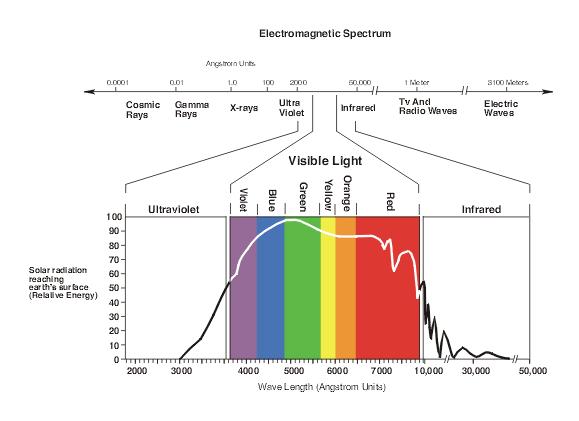
Source: http://www.perret-optic.ch/optometrie/Vision_des_couleurs/vis-couleur_gb.htm
The Eye
Focusing an image
______________ - contractions of ciliary muscles to deform the lens and change the focus
Optional video: Children that can voluntarily control their pupils
http://www.youtube.com/watch?v=YIKm3Pq9U8M

Distance cues from eyes
_________________________ - eyes turn slightly inward for closer objects_________________________ - the eyes have a slightly different perspective
the closer an object, the more obvious the difference in perspective
Saccades
Saccade - quick shifts in eye direction to observe a scenenot the same as the slow tracking when following an object (a car crossing in front of you)
 Source: http://en.wikipedia.org/wiki/Eye_tracking |
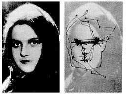 Source: http://www.androidblues.com/visualperception.html |
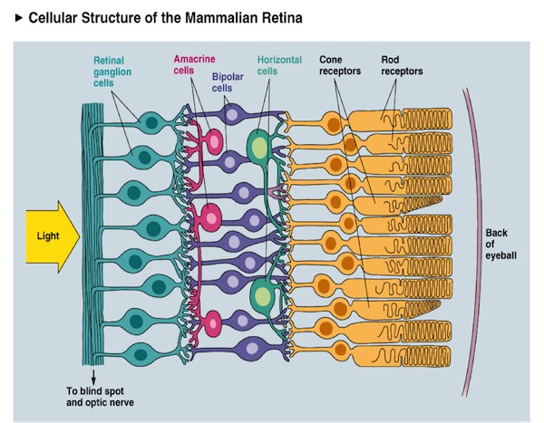
Retina
Transduction
transduction - conversion of one form of energy into another
visual transduction - turning _____________ into a _____________
_____________
how does this happen: pigment absorb photons and react
Rods & Cones
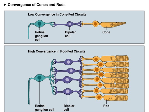
________________________ - cells specialized for visual transductionrods - specialized for seeing ________________________
more sensitive to photons than cones
signals from many rods are pooled into one retinal ganglion cell
cones - specialized for seeing________________________ (more later)
in most humans, there are 3 different cones sensitive to 3 different wavelengths of light
|
|
 |
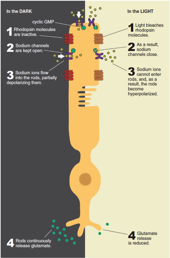
Rhodopsin - a ______________ that changes shape when it absorbs ______________
(you won't be tested on cyclic GMP, just rhodopsin in general:)
cyclic GMP keeps sodium channels open
when rhodopsin absorbs light, it breaks up cyclic GMP
when light hits rhodopsin, this:
increases/decreases the amount of Na+ entering the cell
depolarizes/hyperpolarizes the cell
increases/decreases glutamate release
 |
Rhodopsin: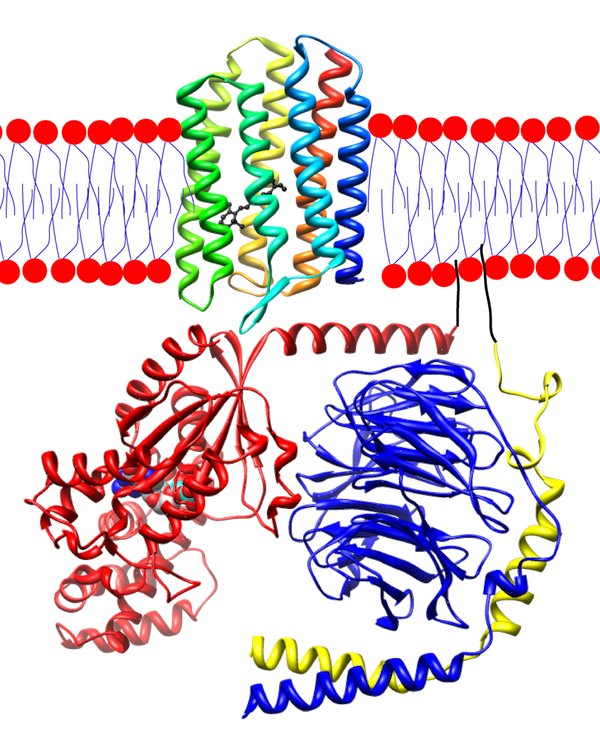 Source: wikipedia.org/wiki/Rhodopsin |
_________________ - the ability to see when light is dim, requires _________________ photoreceptors
_________________ - the ability to see details (resolution), requires ________________ photoreceptors
Other retinal cells
Bipolar cellstypically only connect to a few rods or cones (never both)
"BB" Bipolar mnemonic:
OFF bipolar cells - when it's Bright, off Bipolar cells are off
ON bipolar cells - when it's Bright, on Bipolar cells are on
(reminder: when it's Bright, rods/cones are off)
ON bipolar cells reverse the signal from rods/cones
rods/cones _________________ glutamate release when they absorb light
ON bipolar cells _____________________ to glutamate (WTF?!?)
bright -> rods/cones hyperpolarize -> less glutamate -> less hyperpolarization -> depolarize
Amacrine / Horizontal cells - involved in lateral inhibition (more later)
typically connect to many rods/cones
Retinal ganglion cells - carry the signal from retina out of the eye
may receive input from only a few cones or many rods
their axons form the optic nerve
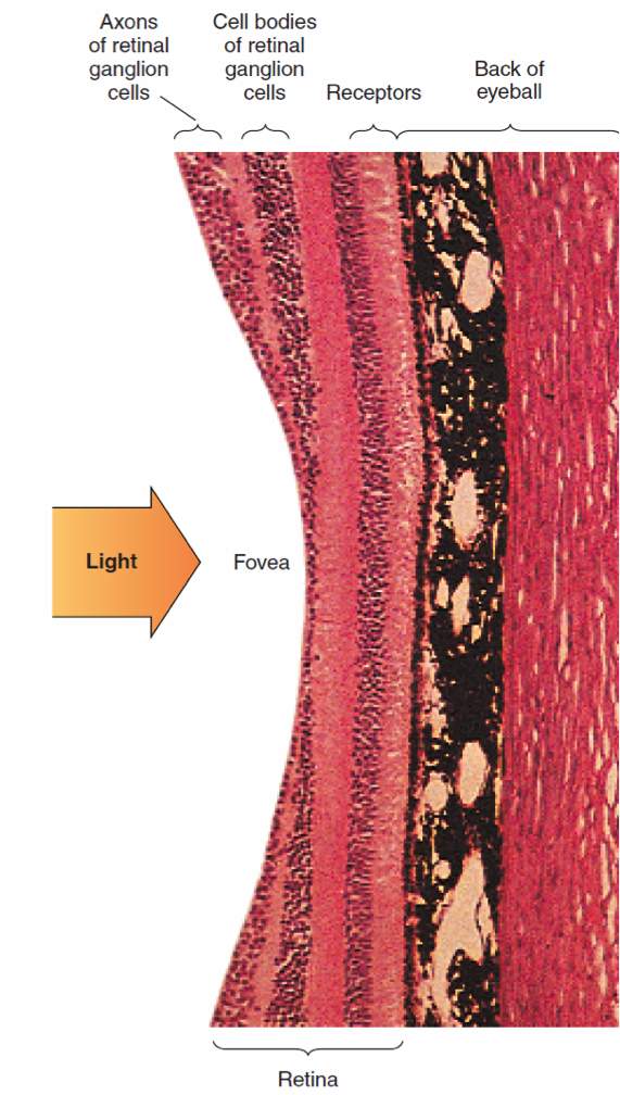
Distribution of rods and cones in the retina:
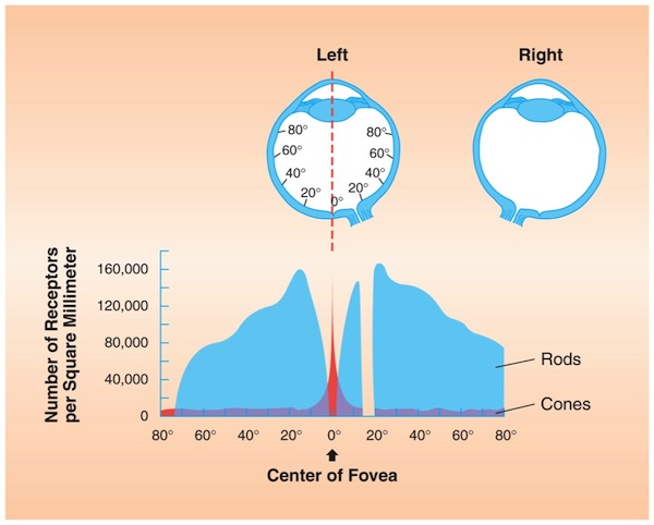
_________________ - the center of the retina
where the lens focuses the image
a high/low concentration of cones
a high/low concentration of rods
_________________ - the area of the retina where the axons from the retinal ganglion cells leave the eye
Trick for seeing in the dark - don't look directly at what you want to see.
Why does this work?
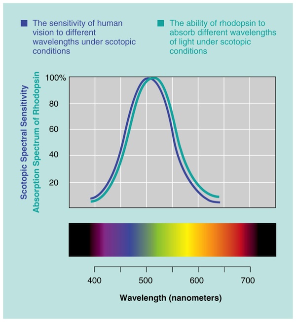
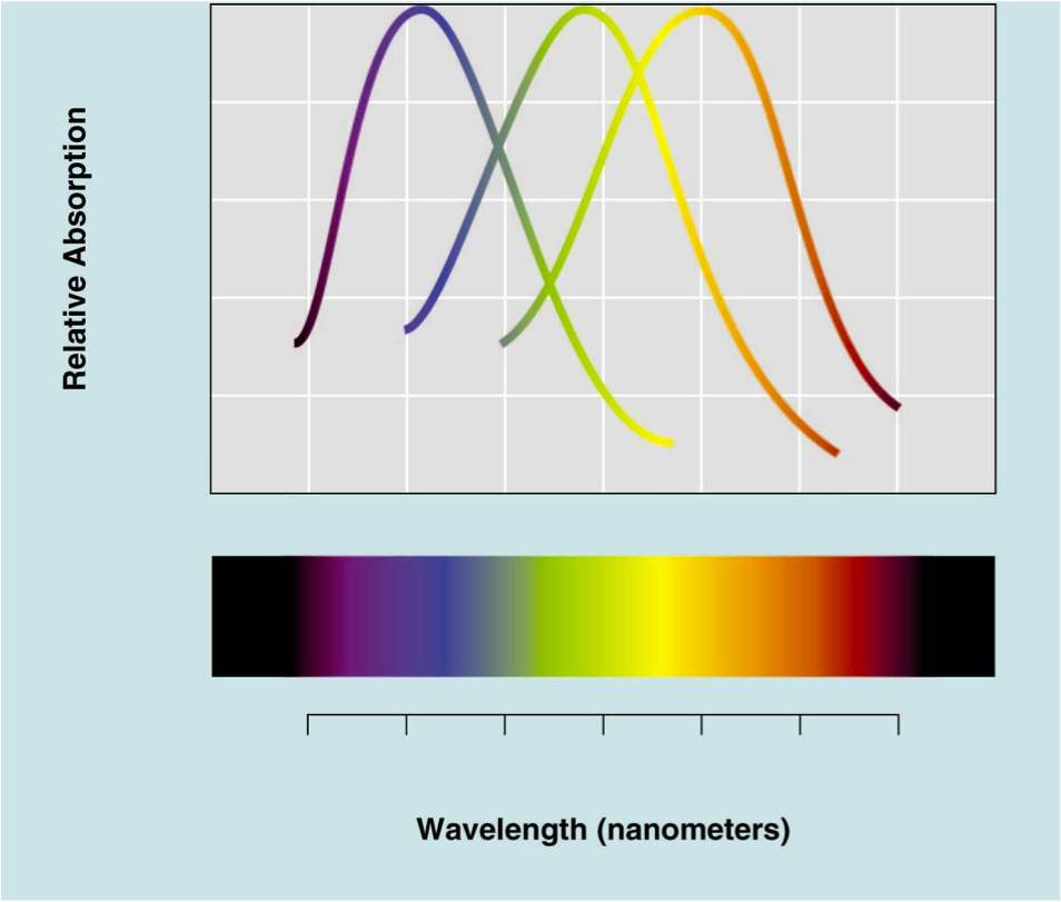
Spectral sensitivity
- pigments will absorb more light from some wavelengths than for others
- spectral sensitivity = a profile of absorption/reaction across different wavelengths
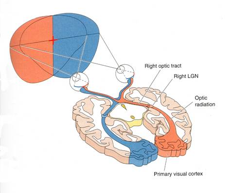
Visual pathway
Source: http://www.dgward.com/physo101/sm06_pages/labs/Peripheral%20Vision%20and%20Visual%20Pathways.htm
Lateral geniculate nucleus (LGN)
- part of the thalamus, which is a relay station between most sense systems and the cortex
- exact role is unclear
- maybe involved in: making visual information more efficient, focusing attention, saccades
Visual cortex (more later)
- performs the processing on visual information to allow us to perceive visual scenes/stimuli
Information from LEFT visual field goes to RIGHT visual cortex (and vice versa)
NOT left EYE to right visual cortex
Retinotopic mapping
- If two retinal ganglion cells that are close together in the retina, their axons end close together in the visual cortex
- The retina is "mapped on" to the cortex
M & P channels
- two parallel channels of axons running through LGN
- magnocellular layers (magno=big, M layers) = movement, "big picture"
- parvocellular layers (P layers) = color, detail, stationary/slow tracking
- demonstrates principle of parallel processing / functional segregation (more later)
Low-level Visual Processing
"low-level" refers to early in the visual pathway & dealing
with simple visual stimuli like brightness, edges & color.
"high-level" refers be areas that receive the pre-processed
information from low-level parts of the visual system and that
process more advanced stimuli like motion, faces,
object-recognition & visual space
Receptive Fields
Definition: The area of visual space that stimulates or inhibits a neuron (or neural tissue)
The stimulus might be simple or complex. Examples:
- Some neurons might be stimulated by any light in a
precise spot in the top right corner of the visual field
- Some neurons might be stimulated by a vertical edge
anywhere on the left
- Some neurons might be stimulated by faces anywhere in
the visual field
Receptive fields become larger
farther away from the fovea - don't need to
know exact location, just want to notice something
at higher levels of the visual system - just
want to react to a face (for example), doesn't matter where it
is
Hubel & Weisel
Videos: Intro
& long
version
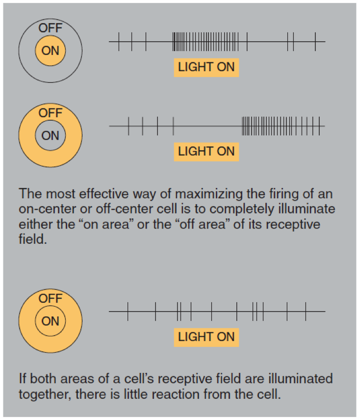
Center-surround
A simple but common kind of receptive field: Center-surround
Behavior:
is _______________ by light in the middle of receptive field
is _______________ by light surrounding the
middle of receptive field
Result:
is most active if it is the only field
receiving input
is not very active if it is receiving input
along with all surrounding areas
increases contrast
uses lateral inhibition
Edges
Why do edges appear "accented"?
Source: http://www.vdic.com/casedatabase/casedatabase_visual_perception.asp
Lateral Inhibition
lateral inhibition increases
contrast between strong and weak signals
When a neuron fires, it inhibits its neighbors
Source: http://www.d.umn.edu/~jfitzake/Lectures/DMED/SensoryPhysiology/GeneralPrinciples/LateralInhibition.html
Color
In most humans, there are three kinds of cones- each with a different photo-sensitive pigment called iodopsins
- each of the three iodopsins is sensitive to different wavelengths of light
Number of cones varies
- some animals & people (with color blindness) have only 2 kinds of cones
- some animals (birds in particular) have 4 kinds of cones
(Do not need to know: component/trichromatic theory or color constancy)
High-level Visual Processing
(content for this section is
provided in Brad's presentation)
Visual cortex
Dorsal vs Ventral stream
Face recognition
Damage & Pathologies
- At the level of retina
- In primary visual cortex
- In secondary visual cortex / association cortices
Principles of Visual Processing
- Parallel processing
- Hierarchical
- Functionally segregated