PSYC 2: Biological Foundations - Fall 2012 - Professor Claffey

Notes: Organization

Version:
10/30/12 - original version

Anatomy of the Nervous System

Content covered in Hans's lecture:
CNS & PNS
Directions/Planes
Protection
Spinal Cord
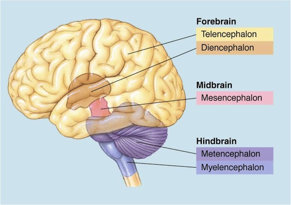
-Encephalonic divisions ( mnemonic: "Start at the Top with T, then alphabetically")
brain divisions

Cerebral cortex

Cortical layers

cortical layers 
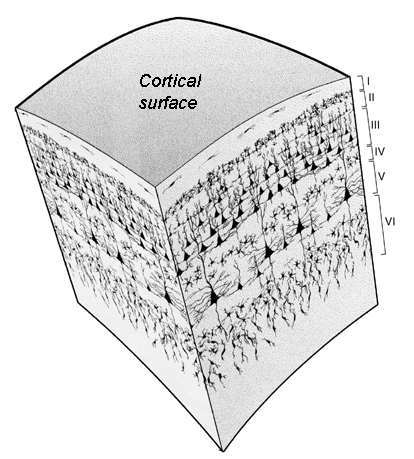
2-4 mm thick with white matter underneath
cortex has layers that differ in neuron organization
some layers consist mostly of:

    _________________ from _____________neurons__
         signals arriving from other areas of the brain (__________)

    ____________________
        densely packed neurons with many synapses (_____________)

    __________________ of neurons whose
        axons project to other cortical areas (__________________)
__________________ cells
    one of the main types of neurons in the cortex
    large multipolar (many extensions) neurons
    many dendrites extending up towards surface of cortex
    large axon that extends down and then to other areas of the cortex
        these axons are what make up the white matter
    integrates signals and communicates to other areas of the brain

Occipital Lobe

Vision (covered in unit 2)

Parietal Lobe

Somatosensory cortex (covered in unit 2)

Association cortex (covered in unit 2)
    integrating vision (dorsal stream) / hearing / touch
    attention
    sense of space and our bodies relation to space
   

Temporal Lobe

(know functions but not names)
superior temporal gyrus (top) - __________________ and __________________

inferior temporal gyrus (bottom) - "what" or ventral stream of __________________

medial temporal lobe (inside) - hippocampus, declarative __________________

Frontal Lobe

primary motor cortex (__________________ movement control)

secondary motor areas (__________________ movement control)

prefrontal cortex - advanced __________________ functions, __________________
    "pre" meaning before or in front of

Subcortical Structures

"subcortical" is everything under the cortex (the very outer surface)
includes areas introduced above: thalamus, hypothalamus, medulla, pons, cerebellum
includes the limbic system (more detail in Unit: Cognition) & basal ganglia (more detail in Motor System below)

Limbic system

limbic system 
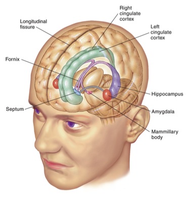
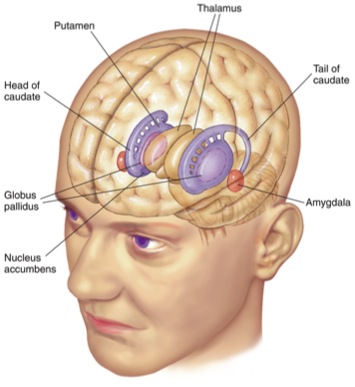
circles the thalamus (limbic is Greek for ring)
regulates the four Fs of behavior:
   fighting, fleeing, feeding, and ______________

Areas

________________
    declarative memory
        knowledge that can be declared as oppose to procedural memory

________________
    (Greek: almond)
    emotional learning, fear & aggressive behaviors


Basal ganglia

To be covered in Motor System of this unit

Motor System

Optional TED Talk - Daniel Wolpert: The real reason for brains (first 2:30 is most relevant, but good stuff after)
   http://www.ted.com/talks/daniel_wolpert_the_real_reason_for_brains.html

Overview
    Responsible for controlling our __
______________/skeletal muscle

    Capable of extensive ________________ and ________________

    Requires extensive somatosensory ________________

Pathways

motor pathways

Upper Motor Neurons

Cell bodies in primary motor cortex
Travel down the spinal cord

Lower Motor Neurons

Synapse onto muscles - neuromuscular junctions
Their action potentials cause muscles to contract

Dorsolateral Pathway

Necessary for ____________________________
Controls the distal muscles (e.g. hands, wrists, feet)
Axons go to only one specific segment of the spine
Lesions cause problems with fine movement (like moving some fingers but not others)

Ventromedial Pathway

Necessary for ________________ and "anti-gravity"
Controls the proximal muscles (e.g. the trunk, neck, chest)
Axons go to many segments of the spine
Lesions cause problems with posture, sitting up, orienting

Brain Areas

motor cortex
motor pathways

Primary Motor Cortex

secondary motor cortex

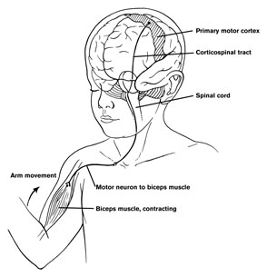
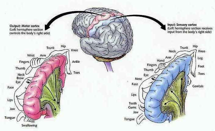
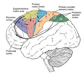
Role: Main source of output signals to control muscles
Input
: secondary/supplementary motor areas &
   somatosensory cortex
Output: spinal cord (motor pathways)

Similar to somatosensory cortex
    Organized somatotopically
    Body regions with more refined control (hands, mouth)
        have larger representations

Electrical stimulation causes:
    Old view:          simple, single muscle contractions
    Recent view:    stereotypical, sometime multi-muscle
                                movements
                            movement towards a given endpoint
                                regardless of starting position

Secondary Motor Cortex

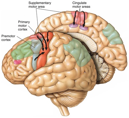
Role: Planning and coordinating complex movements
Input: dorsolateral prefrontal cortex
Output: primary motor cortex

Includes: supplementary motor area and premotor area

Dorsolateral Prefrontal Cortex (DLPFC)

motor parietal pathways

Role: ________________ which movement to make
Input: Posterior parietal
Output: Secondary motor areas

DLPFC is involved in far more than motor control
    Responsible for higher cognitive functions:
        working memory, rules, prediction

Posterior Parietal Cortex

(previously introduced in Unit: Sensation)
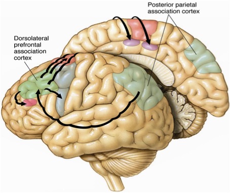
Role:
Representing sense of body and space
Input
: sensory systems
Output: dorsolateral prefrontal cortex (DLPFC)

Damage here can cause problems with voluntary movement (though not habitual/automatic movement)

Basal Ganglia

basal ganglia
Role: Suppress ____________________________ action
Input: cortex
Output: cortex via thalamus

Serves as the gateway for selecting movement
   It is as though the cortex is "proposing" many
   different actions and the basal ganglia selects
   only one

May also select ______________ and ________________ decisions

Cerebellum

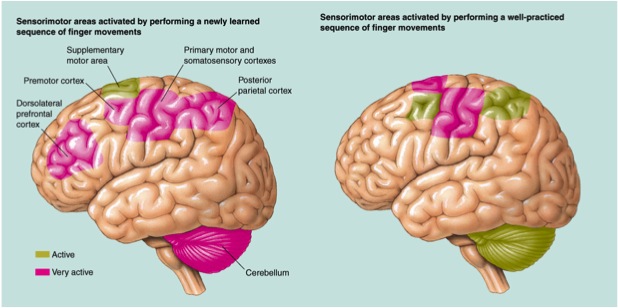
Role: Motor learning and fine coordination
Input: Primary/secondary motor, sensory systems
Output: Primary/secondary motor

Receives a copy of the motor command, compares to desired output, makes small adjustments
Also the site of some motor learning (what people call "muscle memory")


Learning

motor learning
Practice of motor programs changes which areas are involved
Strongly reduced involvement with practice: parietal, DLPFC
Always involved: primary motor cortex, cerebellum, basal ganglia
Practice is relieving higher brain areas of the need to be involved, allowing ________________

Reflexes

motor reflexes 
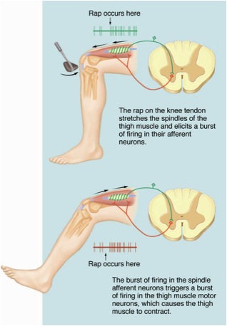
There are sensory neurons that synapse directly on lower motor neurons

Capable of producing movement without the brain

Advantages: ________________ and ________________

Respond to temperature, joint displacement, pressure

Lateralization

________________ - function is more dependent on one hemisphere (left/right) than the other
contralateral - across/different sides
ipsilateral - same side

Lateralization

________________
    problems with speech
    typically patients with aphasia have damage to ________________ hemisphere
    handedness
        right handed people almost always have language in left hemisphere
        left handed people usually have language in the left hemisphere

lateralization language areas
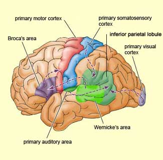
Source: mybrainnotes.com/memory-language-brain.html
Fun Facts (will not be tested)

Paul Broca & Carl Wernicke
 - physicians in 1800's
 - performed autopsies on people with aphasia
 - noticed the reliability of damage to left hemisphere
 - earliest evidence for lateralization in the brain

Wada Test
    anesthetize one hemisphere of the brain at a time
    can impair speech in conscious subjects
    used to localize language before brain surgery

Optional videos:
Broca's aphasia - old, recent
Wernicke's aphasia - old

________________
    problems initiating movement out of context
        movements in context can be routines that don't rely on cortex
    associated with damage to left hemisphere

____________________________
    ignoring one side of the body / space / objects
    typically associated with damage to right parietal lobe

other capabilities
    findings of lateralization for: reading, faces, emotions, music, math, spatial reasoning, details/gist
    often exaggerated in the popular media
    differences can be minor and/or unreliable from person to person
    after damage, other hemisphere can sometimes compensate or develop
    expertise can exaggerate differences

Split-brain patients

 ____________________________ can be surgically cut
    done either experimentally (animals) or to treat epilepsy (humans)
    effects in human patients are obvious/subtle

In cats (Myers & Sperry)

cat split brain 
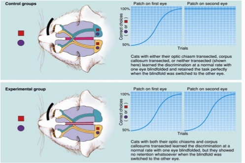
Full description of the experiment on P415-416 (8th ed.)

Four different treatments in the experiment:
A. no surgery
B. cut the optic chiasm
C. cut the corpus callosum
D. cut both optic chiasm and corpus callosum

One hemisphere learned as fast as both hemispheres still connected

Learning could be transferred across corpus callosum

In humans (Gazzaniga)

split                  brain patients: saying versus speaking
Source: brainmind.com/Brain3.html
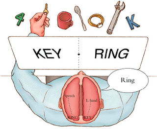
Patients have corpus callosum cut as epilepsy treatment

Different objects/words could be presented to each hemisphere

Person would answer different depending on which hemisphere was responding (remember: language only on left)

2 hemispheres are functioning independently within a person

Videos: Gazzaniga w/ Alan Alda, Other

Development

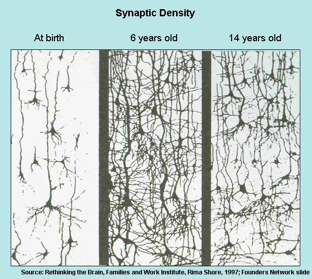
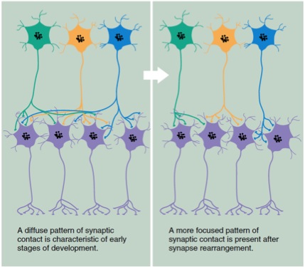
How do we get from embryo to mature adult brain?

Neural Development

1. neural differentiation
    neurons and glia develop from ________________

2. neural proliferation - creation of many new neurons
    happens at the borders of the ventricles

3. neural migration - neurons (without axons or dendrites) spread out to appropriate locations
    guided along paths by glial cells
    aggregate into cohesive structures

4. axon growth
    axon originates from cell body and is able to "find" targets throughout the brain
    growth cone
    chemoaffinity hypothesis - axons are guided by complex chemical signals

5. ____________________________
    growth of new synapses
    dependent on astrocytes

6. ________________
    we lose about 50% of the neurons that are originally created
    apoptosis - programmed self-elimination (cell death)
    neurons start with many more connections than needed,
        then lose connections with other neurons,
        but increase synaptic strength with the connections that remain
        example: initially many motor neurons connect at each neuromuscular junction,
            but in adulthood only one motor neuron remains connected (competition)

development pruning
development focusing
(Timeline)

Timing of Development

24 days - neural tube
40 days - initial swellings of fore/mid/hind brain

maximum neurons at ___ years old

primary visual cortex
    synaptogensis in 4th month post-natal
    "maximum synaptic density (150% of adult levels) in 7th month post-natal"

brain increase 4x in volume from birth to adult hood
    NOT due to ________________
    due to:
        synaptogenesis
        myelination of axons
        increased branching of dendrites

prefrontal cortex
    longest period of development (________________)
    maximum # of neurons in 2nd year
    spurt of neuronal growth just before puberty, then a pruning in adolescence (Giedd 2004)
        losing 1% of gray matter per year
        teens have less myelin than adults in PFC (Giedd 2004)

Critical Periods

critical period - a time when an organism must have an experience to develop a capability
driven by the competitive nature of neurons
examples: visual development (below), imprinting in ducks & geese, necessity of early social experience in primates

sensitive  period - an experience is highly influential on development of a skill
    these tend to be more loosely defined and arguable than critical periods

The effect of depriving one eye on the development of the visual cortex

vision deprivation setup
vision deprivation cortex cartoon
vision deprivation infant adult
vision deprivation 3 vs 6 days
Source: Originally from "Neuroscience" by Purves, posted on bbs.stardestroyer.net/viewtopic.php?t=124049

Principles

Neural development milestones are spread out from 1 month after conception to 20 years old
Neural organization is characterized by upfront overgrowth followed by pruning
Neurons show competitive behaviors during development
Different brain areas have different growth and critical periods


Copyright 2012 - Michael Claffey